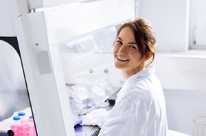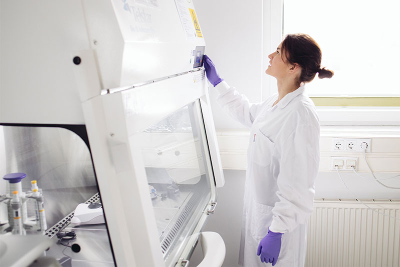
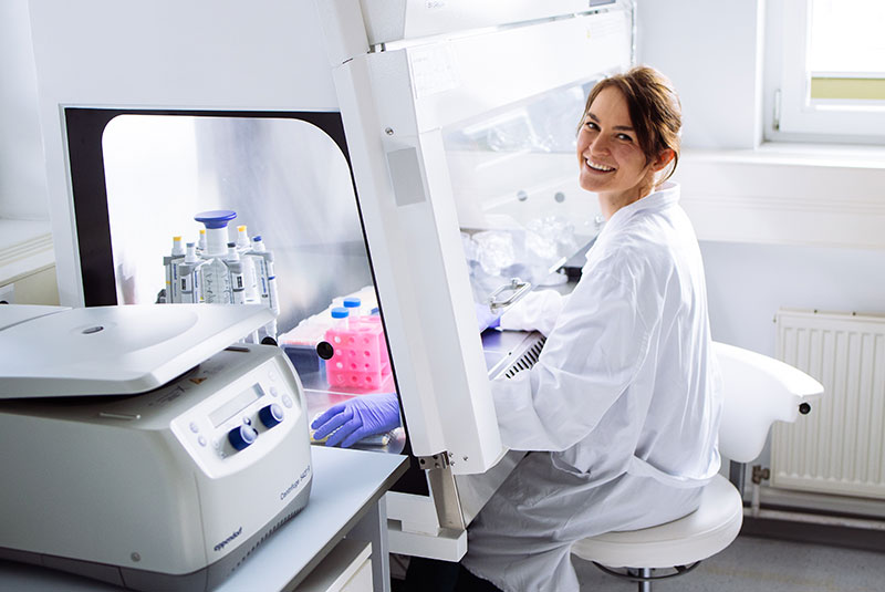
In molecular biology, a safe and clean lab environment is absolutely paramount and the foundation for reliable results. For NGS workflows, and specifically RNA sequencing, the laboratory environment, equipment, and reagents should be kept free of contamination to avoid degradation of the sample through RNases or compromising results through contamination with DNA.
Key Points to Maintaining a Safe and Clean Laboratory Environment and Good Scientific Practice
- Separate laboratory areas to prevent cross contamination, e.g., culturing and sample collection should be separated from labs where molecular assays are run. For PCR-based and NGS assays, adhere to separation of pre- and post-PCR laboratories to avoid contamination of assay material (e.g., sample, library prep components, or equipment) with PCR products (amplicons).
- Unidirectional process: Follow the workflow of each area to avoid contamination during molecular procedures, e.g., always move your samples from pre-PCR to post-PCR labs, but never in the opposite direction; always change gloves and if necessary, also lab coats and other protective gear.
- Ensure your laboratory environment, equipment, and reagents are suited for your experiment, e.g., when working with RNA, ensure the surroundings and materials are free from RNases (see Lexogen’s General Guidelines for more details).
Tip: RNases are ubiquitous and omni-present in all environments. Ensure to clean your workspace with 80 % ethanol and / or RNase removal solutions before starting any RNA assays. As RNase contamination can come from the experimenter, wear gloves (to protect your sample) and avoid speaking over opened tubes. Use sterile and RNase-free materials (pipette tips, plasticware) and RNase-free reagents.
- Use appropriate controls to check your workflow, experiments, and aid with the interpretation of results. Controls ensure the experiment is carried out correctly and the results are useful, e.g., always include positive and negative controls. Include the evaluation of appropriate controls during the experimental planning phase (also see our RNA Lexicon Chapter on Experimental Planning).
- Ensure that functional checks on instruments and equipment maintenance are conducted periodically, e.g., check the functionality / temperature profile of PCR cyclers and ensure your pipets are calibrated.
Tip: Pipettes lose accuracy over time and should ideally be checked approx. 1x per year and re-calibrated if needed (depending on the use). You can also use visual inspection of the volumes taken up by the pipette to have a quick check on the accuracy – several pipette tips, especially the ones used for low volumes 1- 10 µl have specific gradings on the tip.
- Propper pipetting technique also helps to prevent contamination! Ensure the pipette tip is securely fastened on the pipette. Aspirate at a vertical 90° angle with consistent pressure and speed. For dispensing, the pipette should be held at a 45° angle. Ensure not to touch the insides of reagent tubes with the pipette.
- Perform laboratory environmental monitoring to meet equipment specifications (e.g., humidity, temperature). This is especially important for sensitive instruments such as sequencers.
- Monitor refrigerators and freezers containing any samples or reagents to prevent samples from spoiling due to power loss or equipment failure.
- Do not use expired reagents or reagents stored at the wrong temperature, functionality of the assay and the results can be compromised.
- Make sure that there is enough space so that quality of work, cleanliness of the workplace, and safety of personnel are not compromised. Check these other important points for your lab environment and workspace:
- fire prevention,
- electrical safety,
- chemical safety,
- radiation safety,
- environmental safety, take care to dispose laboratory waste appropriately.

Keep it Clean: Why Separating Pre-PCR and Post-PCR Labs Matters
PCR is extremely sensitive! The Polymerase Chain Reaction (PCR) amplifies minuscule amounts of DNA. Therefore, even a trace of amplified DNA (from a previous experiment) can contaminate new samples or reagents used in the assay falsifying results. To learn more about PCR, see our Lexicon Chapter on Molecular Biology Basics.
Two spaces to keep it clean!
- Pre-PCR = “clean area”: In this area precious samples are handled prior to amplification. Keeping it free of “contaminating” amplified DNA is key for reliable and correct results.
- Post-PCR = for amplified DNA: Once DNA is amplified, contamination risk to the current experiment is no longer a threat. In the post-PCR area, the sample is amplified, and the resulting DNA library is analyzed, e.g., by electrophoresis, Fragment Analyzer, NanoDrop, and further prepared for subsequent sequencing.
Separation can be simple:
- Two rooms: Ideally, pre-PCR and post-PCR labs are situated in dedicated rooms that can be closed individually.
- Dedicated equipment: Use separate instruments (PCR cyclers, fridges, freezers, centrifuges), equipment e.g., pipettes, racks, magnetic separators, and reagents in each area and do not transfer materials to avoid contamination.
- Airflow matters: Slightly positive air pressure in pre-PCR will prevent contamination from the post-PCR area.
- Never move samples, equipment, or reagents from post-PCR to the clean pre-PCR area without thorough decontamination. Always change gloves and ideally protective equipment when moving between areas.
By separating these areas, you ensure the accuracy and reliability of your experimental results, ultimately leading to better science!
Aysal, A. et al., (2020) How to Set Up a Molecular Pathology Lab, Journal of Pathology (36) 179-187, doi: 10.5146/tjpath.2020.01488
Ou CY, et al., (1991). Use of UV irradiation to reduce false positivity in polymerase chain reaction. Biotechniques (10) 442-446.
Hu Y. (2016) Regulatory concern of polymerase chain reaction (PCR) carryover contamination. In: Samadikuchaksaraei, A, ed. Polymerase Chain Reaction for Biomedical Applications. IntechOpen, 57-68. doi: 10.5772/66294.
Prince AM, Andrus L. (1992) PCR: how to kill unwanted DNA. Biotechniques. (23) 358-360.
Mifflin TE. (2007) Setting up a PCR laboratory. CSH Protoc. pdb.top14.
Written by Dr. Yvonne Goepel
Image courtesy of Mag. Amra Dedic